


If your child is seeing an orthodontist, you may hear the words “we’ll take some X-rays” early in the process. For many parents, that raises two immediate questions: “Are dental X-rays safe for kids?” and “What exactly is the orthodontist looking for?” Both are valid, and the answers are reassuring when you understand how modern orthodontic imaging works.
Dental X-rays are diagnostic tools that help orthodontists see what the eyes cannot. They reveal developing teeth under the gums, jaw growth patterns, airway and bone structures, and bite relationships that guide treatment decisions. In pediatric orthodontics, X-rays can help catch issues early, plan treatment more precisely, and avoid surprises later.
This guide explains the most common orthodontic X-rays used for children, what they show, how safety is managed, and how often imaging is typically needed. If you want a broader view of how imaging fits into early care, start with the overview of orthodontic services.
Why orthodontists use X-rays for children
A child’s mouth is changing fast. Between ages 6 and 13, permanent teeth erupt, roots finish forming, and jaw growth accelerates. Many orthodontic problems develop quietly during this stage. Even if teeth look “pretty straight” on the outside, there can be hidden concerns such as:
- Permanent teeth that are delayed, missing, or erupting in the wrong direction
- Crowding developing under the gums before it is visible
- Bite discrepancies caused by jaw growth differences
- Extra teeth, unusual root shapes, or other developmental variations
- Teeth that may become impacted, especially canines
X-rays help orthodontists make decisions based on structure, not guesswork. This is one reason early evaluations can be so valuable. If you are learning about timing, the general guidance on early orthodontic planning is often discussed during a first visit, and you can request an evaluation through Contact Us.
The big safety question: Are dental X-rays safe for kids?
Parents are right to think about radiation exposure. The good news is that orthodontic offices use modern imaging systems designed to keep exposure low and only take images when there is a clear diagnostic benefit.
Pediatric orthodontic imaging follows a simple principle: take the right image at the right time for a clear clinical reason. Orthodontists do not take X-rays “just because.” Imaging is chosen based on your child’s age, growth stage, and the specific question the orthodontist needs to answer.
How orthodontic practices reduce exposure
While each office has its own protocols, safety practices commonly include:
- Digital imaging that uses lower exposure than older film systems
- Fast sensors that capture the image quickly
- Collimation and proper positioning to limit the area imaged
- The fewest images needed to make a confident diagnosis
- Avoiding repeat images unless clinically necessary
If you want insight into how a practice approaches technology and patient care decisions, you can explore What Sets Us Apart.
What types of dental X-rays are used in pediatric orthodontics
Orthodontic imaging is not one size fits all. Different images answer different questions. Your child may need one type of X-ray, several types, or none on a particular visit.
Below are the most common imaging types used in pediatric orthodontics and what each one helps diagnose.
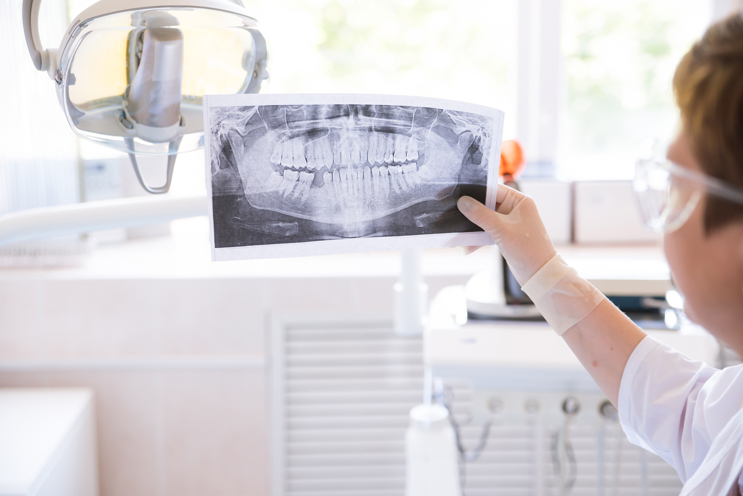
Panoramic X-ray: the full picture of teeth and jaws
A panoramic X-ray is a single image that captures the entire mouth, including:
- All teeth, including those still under the gums
- Upper and lower jaws
- Jaw joints (TMJ area)
- Sinus regions near the upper jaw
What orthodontists look for on a panoramic image
This type of image is often used to evaluate:
- Missing teeth (congenitally absent teeth)
- Extra teeth
- Delayed eruption patterns
- Impacted teeth
- Root development
- Bone levels and general dental health
For children, the panoramic image is especially helpful because it shows both baby teeth and developing permanent teeth at once. It helps orthodontists understand whether spacing issues are likely to resolve naturally or whether early interceptive planning may be needed.
Cephalometric X-ray: measuring growth and bite relationships
A cephalometric X-ray (often called a “ceph”) is a side view image of the head that shows:
- The relationship between the upper and lower jaws
- Facial growth patterns
- Tooth positions relative to the jaws
- Airway space in the throat region
Why a ceph matters for pediatric orthodontics
Growth is a major factor in orthodontics. A ceph helps orthodontists evaluate whether a child’s bite issue is mostly dental (teeth position) or skeletal (jaw growth pattern). That distinction matters because it influences timing and treatment options.
Cephalometric analysis can help with planning for cases involving overbites, underbites, open bites, and facial growth imbalances. It also supports more accurate forecasting of how the bite may change as your child grows.
Bitewing X-rays: checking for cavities and bone support
Bitewing X-rays are commonly taken by general dentists, but orthodontists may request recent bitewings if there are concerns about tooth health before starting treatment. Bitewings show:
- Areas between teeth where cavities often form
- The height of bone supporting the teeth
Orthodontic treatment works best when the teeth and gums are healthy. If a child has active cavities or gum inflammation, the orthodontist may recommend addressing those issues before moving forward with appliances.
For tips on daily care during orthodontic treatment, including hygiene priorities that help prevent issues, parents often find Life With Braces helpful, even if your child is not in braces yet.
Periapical X-rays: zooming in on specific teeth
Periapical images show a close-up of one or a few teeth, including the roots and surrounding bone. Orthodontists may use these when they need more detail about:
- Root shape and length
- A suspicious area on the root or bone
- Eruption direction of a developing tooth
- A tooth that has experienced trauma
These are not always needed in routine orthodontic planning, but they can be very useful for specific questions.
CBCT: 3D imaging for complex cases
Cone Beam CT (CBCT) imaging creates a three dimensional view of teeth, roots, and bone. It is not used for every child. Orthodontists generally reserve CBCT for cases where 2D imaging does not provide enough clarity, such as:
- Impacted teeth in tricky positions
- Complex eruption problems
- Significant bite discrepancies
- Surgical planning or advanced interdisciplinary cases
CBCT can be extremely informative because it shows structures in 3D, including root proximity and exact tooth location. The decision to use CBCT should always have a clear reason, and parents should feel comfortable asking why it is recommended.
What orthodontists are diagnosing with X-rays
X-rays are not about “finding problems” to justify treatment. They are about understanding development so orthodontists can choose the safest, most effective plan. In pediatric orthodontics, imaging helps answer questions like these:
1) Are all permanent teeth present and developing normally?
Children can have missing permanent teeth or extra teeth. Either can affect spacing, bite planning, and long term treatment decisions.
2) Is there enough room for the adult teeth that have not erupted yet?
Crowding is often predictable before the teeth appear. Imaging shows whether permanent teeth are stacked, rotated, or blocked.
3) Are any teeth at risk of becoming impacted?
Upper canines are a common concern. If a canine is drifting off course, early intervention may create room and guide eruption.
4) Are the jaws growing proportionally?
Jaw growth differences may influence when treatment should start and what kind of appliances are most helpful.
5) Is there a bite shift or crossbite pattern?
Some children shift their jaw to make their bite feel comfortable. Imaging can help confirm whether the jaw is narrow or whether the bite relationship is off.
6) Are there signs of previous trauma or root concerns?
If a child has had an injury to the mouth, imaging can help confirm whether roots and bone look healthy.
How often are orthodontic X-rays needed for kids?
There is no single schedule that applies to every child. Frequency depends on:
- Age and growth stage
- Whether the child is being monitored or treated
- Whether new teeth are erupting in a way that changes the plan
- Whether a specific concern needs follow up
Many children have imaging at an initial evaluation, then additional images only when needed for treatment planning or progress checks. If your child is in a monitoring phase, the orthodontist may not need new images at every visit.
A helpful approach as a parent is to ask: “What decision does this X-ray help you make today?” A good answer should connect the image to a clear clinical purpose.
What parents should ask before or after imaging
Most parents do not need to become imaging experts, but a few questions can give you clarity and confidence:
- What type of X-ray are you taking, and what will it show?
- Why is it needed today?
- Do you already have recent images from our general dentist that can be used?
- Will this change the treatment plan or timing?
- What are you specifically evaluating in my child’s case?
If your child has a broken appliance, discomfort, or urgent concern while in treatment, your orthodontist may also reference imaging as part of troubleshooting. For immediate care guidance, parents often start with orthodontic emergencies.
Preparing your child for dental X-rays
Most kids do well with orthodontic imaging, especially when you frame it calmly. Here are simple ways to help:
- Explain that the camera “takes a quick picture” and it does not hurt
- Practice standing still for a few seconds at home
- Remind them to follow the assistant’s instructions about biting or head position
- Avoid big promises like “it will be over in one second” and instead say “it is quick and we will be done soon”
- Praise effort and cooperation afterward
If your child is nervous, ask the team to explain each step. Pediatric orthodontic teams are used to helping kids feel comfortable and confident.
How X-rays support better treatment outcomes
When orthodontists have the right images, they can often reduce delays and avoid mid treatment surprises. Imaging supports:
- Better timing for early treatment when needed
- More predictable eruption planning
- More accurate bite correction strategies
- Safer management of impacted teeth
- Cleaner coordination with your child’s general dentist
It also helps orthodontists explain what they are seeing. Many parents feel relief when they can visualize the problem and understand the plan.
If you are exploring treatment options and want a clear overview of what may be recommended, visit Orthodontic Services to see common paths for children and teens.
Common misconceptions about pediatric orthodontic X-rays
“X-rays are only for braces.”
Not true. Imaging is often most helpful before treatment, when orthodontists are evaluating growth and eruption patterns.
“If teeth look straight, my child does not need X-rays.”
Appearance can be misleading. A child can have hidden crowding, delayed eruption, missing teeth, or bite discrepancies that are not visible from the outside.
“More X-rays means better care.”
Not necessarily. Better care means the right images for the right reason, taken only when needed.
“An orthodontist will always take new X-rays even if my dentist has them.”
Not always. Many orthodontists can use recent images from your general dentist if they are clear and current. It is worth asking.
Conclusion
Dental X-rays play an important role in pediatric orthodontics because they reveal what is happening beneath the surface. They help orthodontists evaluate tooth development, jaw growth, bite relationships, and eruption patterns so treatment decisions are accurate and well timed. Modern orthodontic imaging is designed to be efficient and safety focused, using targeted images only when they provide real diagnostic value.
If you have questions about your child’s dental development or you want a clear plan for timing, scheduling a pediatric orthodontic consultation is a smart next step. You can request an appointment through Contact Us and ask the team what imaging, if any, is recommended for your child’s age and needs.
Frequently Asked Questions About Dental X-Rays in Pediatric Orthodontics
1. Are dental X-rays safe for children?
Yes. Modern orthodontic X-rays use low radiation levels, and orthodontists take images only when there is a clear diagnostic benefit for your child’s care.
2. Why does my child need X-rays if their teeth look straight?
Teeth can look straight on the outside while issues develop under the gums, such as crowding, impacted teeth, missing teeth, or jaw growth concerns. X-rays help reveal what cannot be seen during a visual exam.
3. What is the difference between a panoramic X-ray and a cephalometric X-ray?
A panoramic X-ray shows all teeth and both jaws in one wide image. A cephalometric X-ray is a side view that helps evaluate jaw growth, bite relationships, and facial development patterns.
4. How often will my child need orthodontic X-rays?
It depends on your child’s age, growth stage, and whether they are being monitored or actively treated. Many children only need imaging at key planning points rather than at every visit.
5. Can the orthodontist use X-rays taken at my child’s general dentist?
Often, yes, if the images are recent and clear. It is a good idea to bring copies or request that your dentist send them so the orthodontist can determine if new imaging is necessary.
6. What can X-rays tell the orthodontist about my child’s future teeth?
X-rays show developing permanent teeth, eruption paths, root development, and whether teeth are missing, extra, or at risk of becoming impacted. This helps orthodontists plan treatment timing accurately.
7. Will taking X-rays hurt my child?
No. Dental X-rays are painless. Your child may be asked to bite on a small holder or stand still briefly, but the imaging itself does not cause discomfort.
8. What is a CBCT scan and will my child need one?
A CBCT scan is a 3D image used for more complex cases, such as impacted teeth or advanced planning needs. Most children do not need CBCT, and it is typically recommended only when 2D imaging is not enough.
9. Do orthodontic X-rays help detect impacted teeth early?
Yes. X-rays can reveal teeth that are not erupting correctly and may become impacted, especially upper canines. Early detection can allow simpler intervention and reduce future complications.
10. What should I ask my orthodontist about X-rays during the appointment?
Ask what type of X-ray is being taken, what the orthodontist is looking for, and how the image will influence treatment timing or recommendations. A clear explanation should connect the imaging to a specific clinical purpose.